Clinical Diagnostic Parasitology Laboratory (CDPL)
Other diagnostic tests
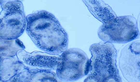
Echinococcus – hydatid
Cyst fluid for Hydatid disease (Echinococcus species) UKAS accredited test
Aspiration of cyst should only be considered after taking expert advice.
Specimen requirement: Cyst fluid should ideally be examined within two days of sample being taken. No minimum sample requirement.
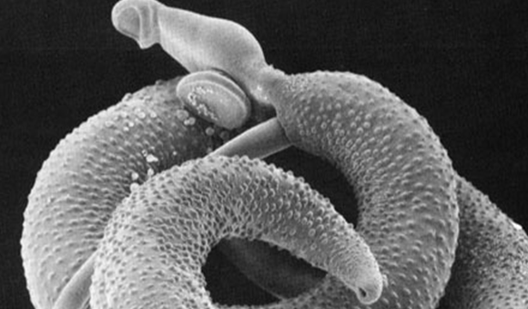
Schistosomiasis
Faecal Parasite diagnosis – UKAS accredited test
Faecal microscopy
A concentration technique is routinely performed on all faecal samples for the presence of cysts, ova and larvae.
Sample requirements:
- Minimum of a quarter specimen pot of faecal sample.
- Ova, cysts, and parasites may be passed intermittently so three samples may need to be examined.
- Ova, cysts, and parasites will diminish over time so it is ideal if the sample is less than two days old upon receipt at LSTM. However, older stools will not be rejected.
Urine techniques – UKAS accredited test
Schistosoma haematobium
Urine is tested by dipstick for the presence of blood Hb, and RBCs. Urine is routinely filtered for Schistosomiasis ova using polycarbonate filters.
Sample requirements:
- Ideally total 10.00-14.00 urine collection or terminal 12.00 sample
Key factors affecting test:
Random urine samples may give false negative results
Semen – UKAS accredited test
Seminal fluid microscopy is performed for schistosome ova.
Specimen requirements:
Age and volume of the sample is not critical.
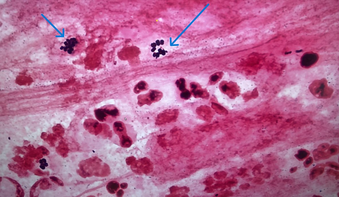
Leishmania
Leishmania diagnosis Microscopy – UKAS accredited test
Sample requirements:
See downloadable sampling notes.
Cutaneous Leishmaniasis
- Unstained, fixed aspirate or biopsy impression smears for microscopy.
- Giemsa-stained histology slides.
- Biopsy material in PCR (ATL) buffer (available from laboratory on request), OR in dry, sterile container OR in 10% ethanol. If histological wax block of tissue is the only material available please cut 10µm thick wax sections and float onto microscope slides (3/slide, two or 3 slides/sample), send these unstained for DNA extraction. A biopsy should ideally be around the size of a grain of rice.
Visceral leishmaniasis
- Thin marrow smears (please fix for one minute in methanol before sending.)
- Marrow /blood in EDTA for PCR -minimum of 500µl.
Key factors affecting test:
Use of aspirate, wax block or samples smaller than the size of a grain of rice may cause false negative or insufficient DNA results. The use of Iodine during sampling will inhibit the PCR amplification and may cause false negative results. The use of Lithium heparin instead of EDTA will inhibit the PCR amplification and may cause false negative results. Please list travel history with all Leishmania PCR requests.

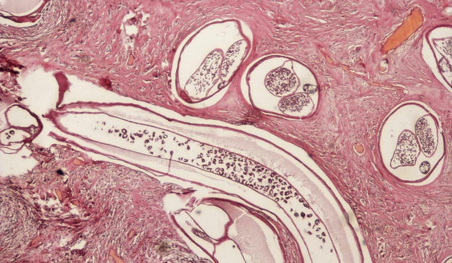
Onchocerciasis
Microscopy for Onchocerciasis – UKAS accredited test
Skin snips can be examined for the presence of microfilariae. Please contact the laboratory before taking the sample from the patient.
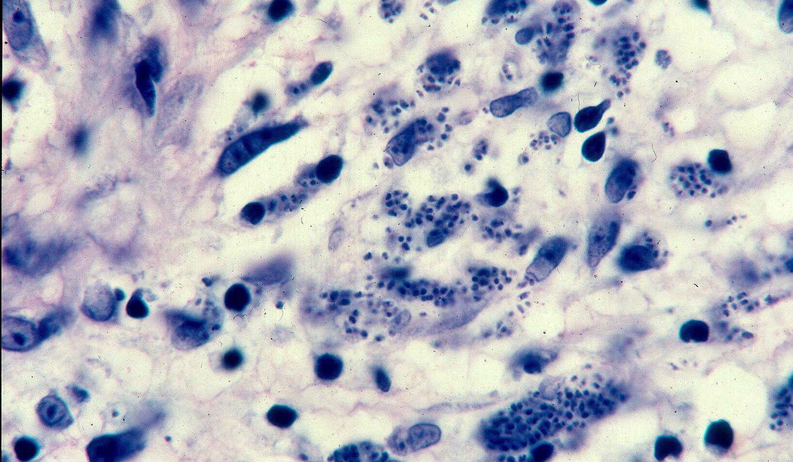
Histology sections
Histology sections should be sent stained for parasite microscopy. Ideally H&E stained sections plus Giemsa stained section if querying Leishmaniasis or a PAS stained section if querying Amoebae.
Examination of sputum
Sputum microscopy is performed for parasitic ova.
Specimen requirements:
- Age and volume of the sample is not critical.
- The laboratory does not process samples when a diagnosis of TB cannot be excluded.