Clinical Diagnostic Parasitology Laboratory (CDPL)
Blood parasites
Malaria
Samples are screened using thick blood films. Thin films are examined on positive samples. If P.falciparum is found, parasitaemia is performed. Rapid diagnostic tests (RDTs) are available.
Sample requirements:
• Thick and thin films (unstained but thin-film methanol fixed) made from a fresh blood sample.
• Thick and thin films made and stained from a fresh blood sample.
• Original EDTA sample.
Key factors affecting tests:
Malaria parasites collected into anticoagulants such as EDTA deteriorate and morphological changes occur within a few hours.
Complete the malaria reference laboratory form and the report form, and enclose them with the sample. Include sample date and patient travel history.

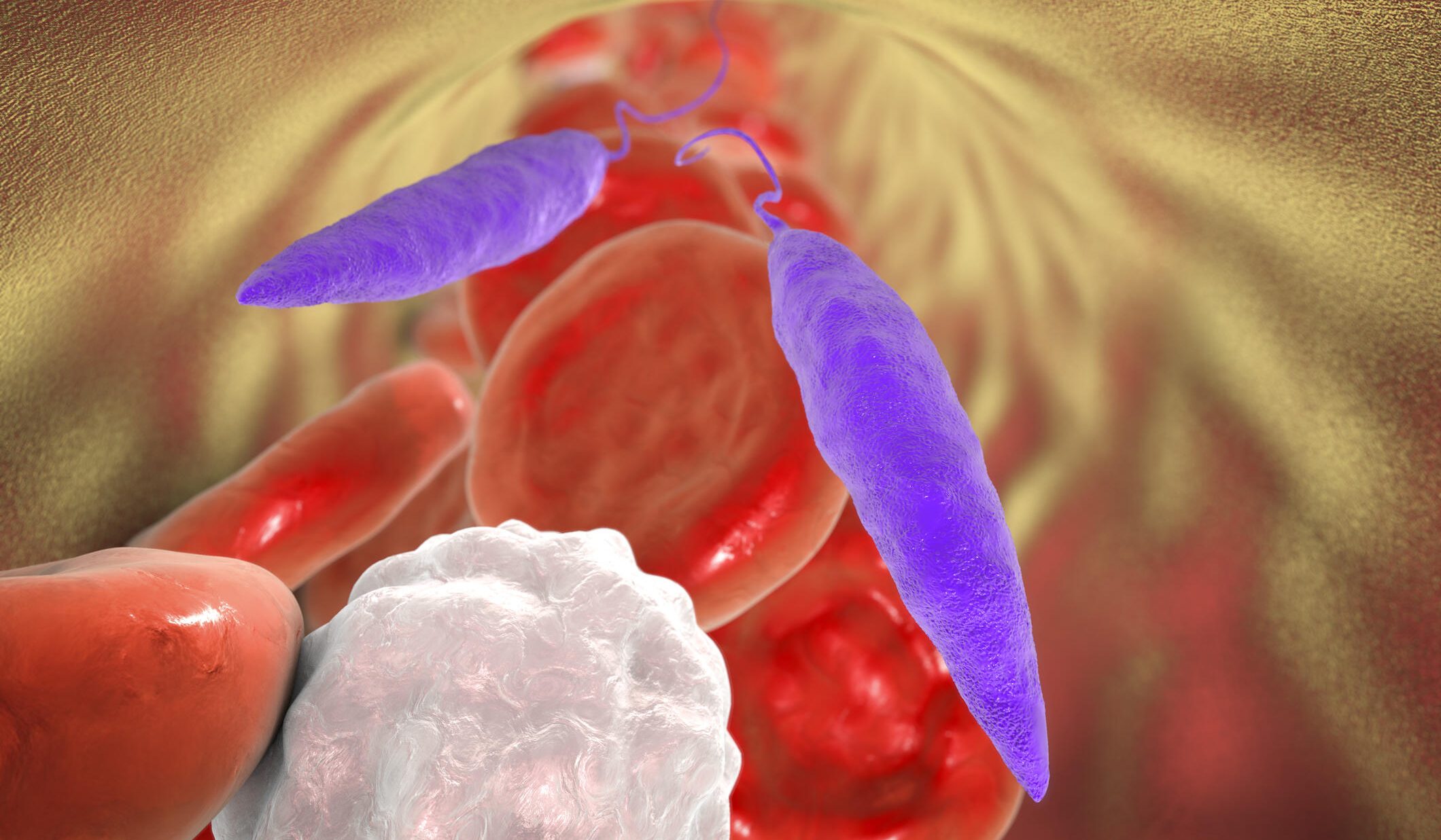
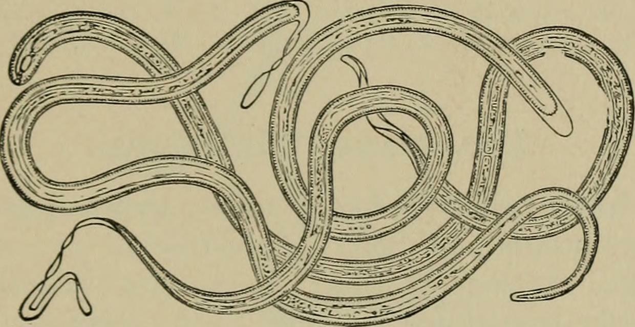
Filaria
A definitive diagnosis of filariasis is usually made by the demonstration of microfilariae in the peripheral blood. The exception to this is Onchocerca volvulus which is diagnosed by demonstration of microfilariae in skin snips.
Blood filariasis:
A wet preparation is examined and samples are filtered using a Nuclepore membrane. If W.bancrofti is suspected, blood collection time should be between 22.00 and 02.00. For a Loa loa diurnal blood sample, 12.00 is preferred.
Sample requirements:
- EDTA blood.
- Sample size is not critical but ideally it should be between 5ml and 10ml.
- The larger the sample filtered, the greater chance of demonstrating the microfilariae.
Key factors affecting tests:
Sheathed microfilariae may ex-sheath if the blood sample is not examined within two to three days of collection. All microfilariae may last up to 72 hours in EDTA blood before disintegration.
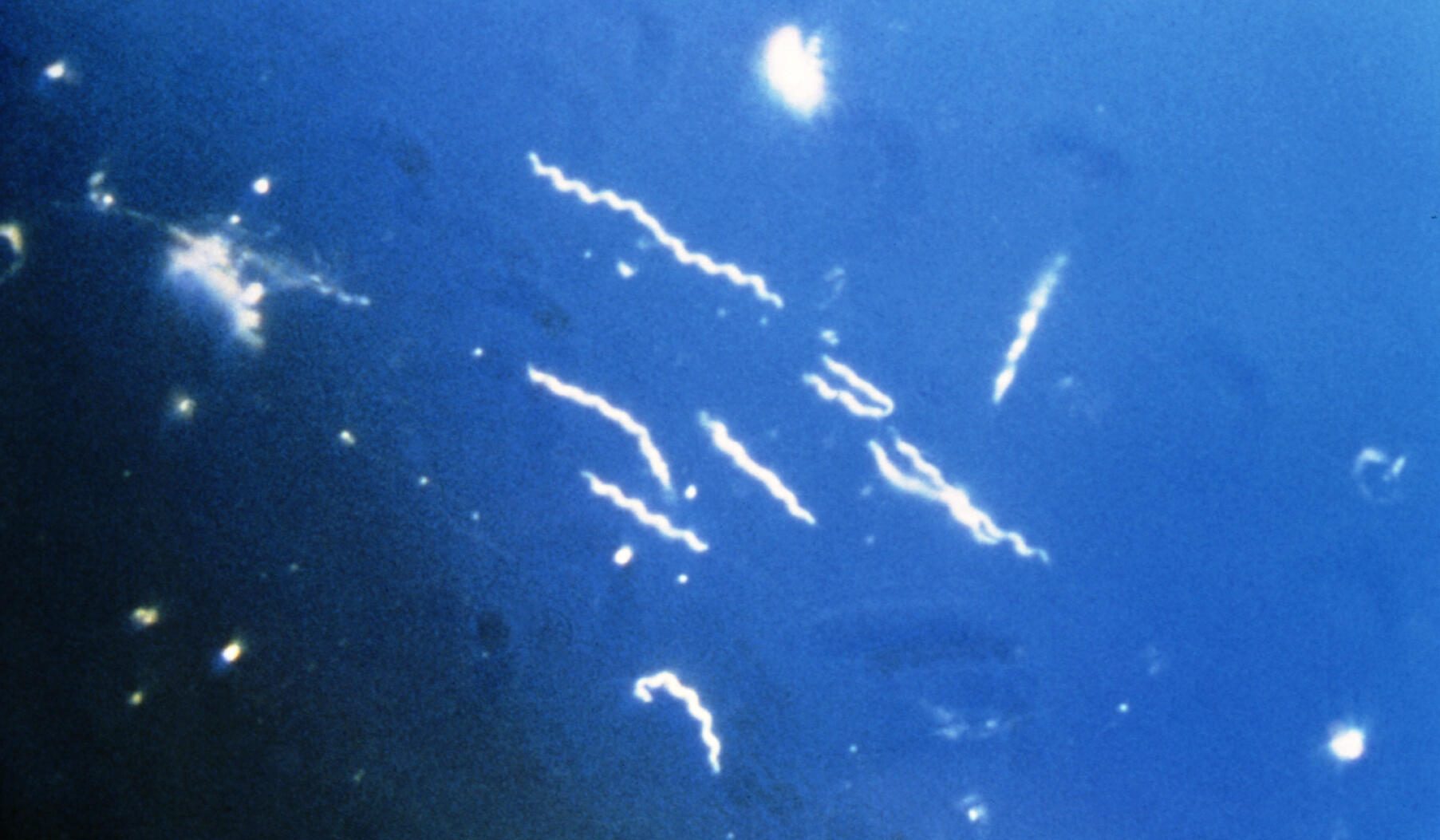
Trypanosomiasis
Thick blood films are used and concentration technique is applied if films are negative. Blood samples for concentration should be examined on the day of collection.
Sample requirements:
- Unstained thick/thin films.
- EDTA blood sample.
Key factors affecting tests:
Trypanosomes may not survive for more than a few hours in EDTA tube, so posted samples can show false-negative results.
If a CSF sample is to be examined for trypanosomes, this should ideally be done within 20 minutes of L.P.

Babesia
Sample requirements:
- Thick and thin films (unstained but thin-film methanol fixed) made from a fresh blood sample.
- Thick and thin films made and stained from a fresh blood sample.
- Original EDTA sample.

Borrelia
Sample requirements:
- Thick and thin films (unstained but thin-film methanol fixed) made from a fresh blood sample.
- Thick and thin films made and stained from a fresh blood sample.
- Original EDTA sample.
Leishmania
Sample requirements:
- Thick and thin films (unstained but thin-film methanol fixed) made from a fresh blood sample.
- Thick and thin films made and stained from a fresh blood sample.
- Original EDTA sample.