Clinical Diagnostic Parasitology Laboratory (CDPL)
Intestinal parasites
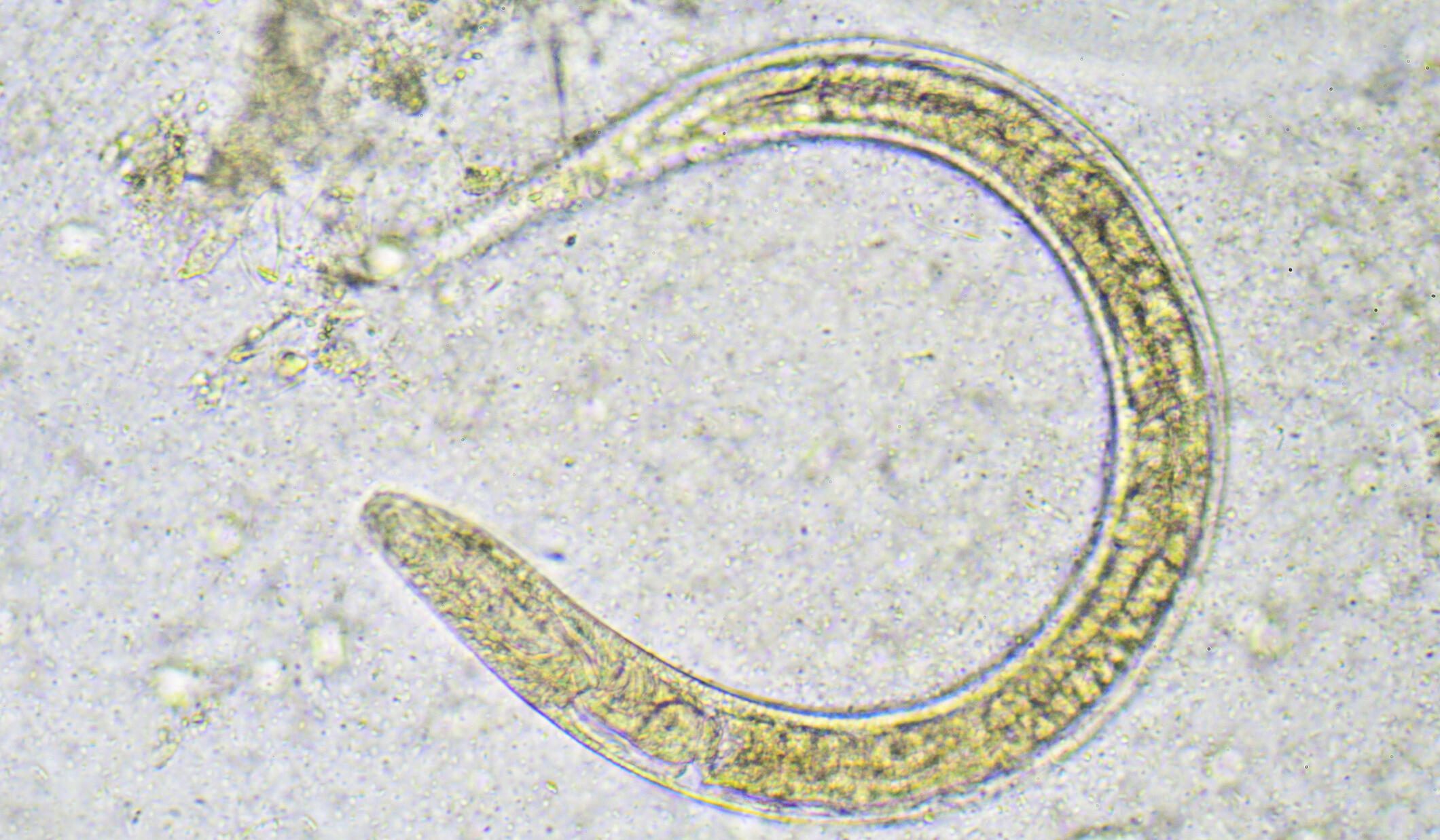
Strongyloides
Faecal microscopy: UKAS accredited test
A concentration technique is routinely performed on all faecal samples for the presence of cysts, ova, and larvae.
Sample requirement:
Minimum of a quarter specimen pot of faecal sample. Ova, cysts, and parasites may be passed intermittently therefore three samples may be required to be examined. Ova, cysts, and parasites will diminish over time so it is ideal for the sample to be less than two days old upon receipt at LSTM. However, older stools will not be rejected.
Culture for Strongyloides/Hookworm: UKAS accredited test
Faeces are cultured for filariform larvae using the charcoal technique.
Sample requirements:
- At least 50g (half pot) of faeces.
- Ova, cysts, and parasites may be passed intermittently so three samples may need to be examined.
Key factors affecting tests:
Samples should NOT be stored in a refrigerator or cold room following collection as this may inhibit subsequent larval growth.
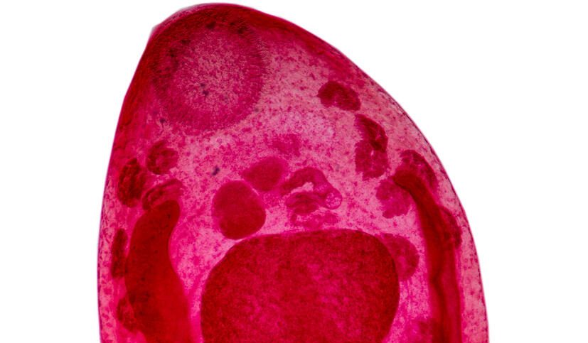
Schistosomiasis
Faecal Parasite diagnosis – UKAS accredited test
Faecal microscopy
A concentration technique is routinely performed on all faecal samples for the presence of cysts, ova, and larvae.
Sample requirements:
- Minimum of a quarter specimen pot of faecal sample.
- Ova, cysts, and parasites may be passed intermittently so three samples may need to be examined.
- Ova, cysts, and parasites will diminish over time so it is ideal if the sample is less than two days old upon receipt at LSTM. However, older stools will not be rejected.
Urine techniques – UKAS accredited test
Schistosoma haematobium
Urine is tested by dipstick for the presence of blood/Hb, and RBCs. Urine is routinely filtered for Schistosomiasis ova using polycarbonate filters.
Sample requirements:
- Ideally total 10.00-14.00 urine collection or terminal 12.00 sample
Key factors affecting test:
Random urine samples may give false-negative results.
Semen – UKAS accredited test
Seminal fluid microscopy is performed for schistosome ova.
Specimen requirements:
- Age and volume of the sample is not critical.

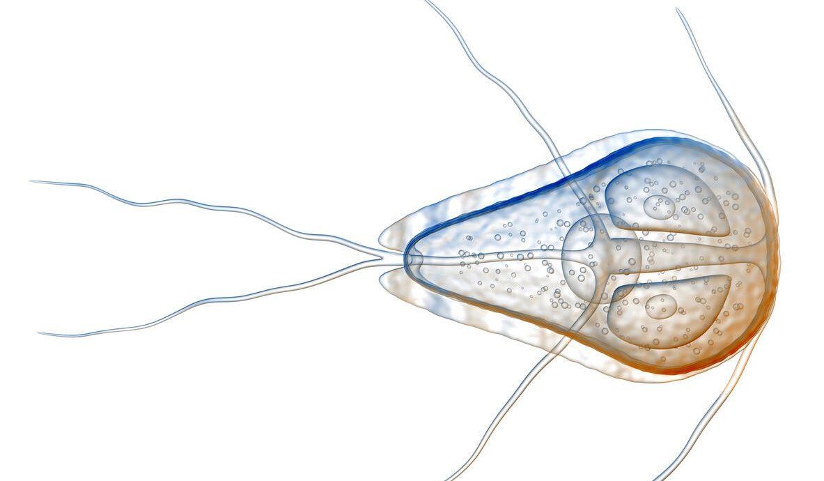
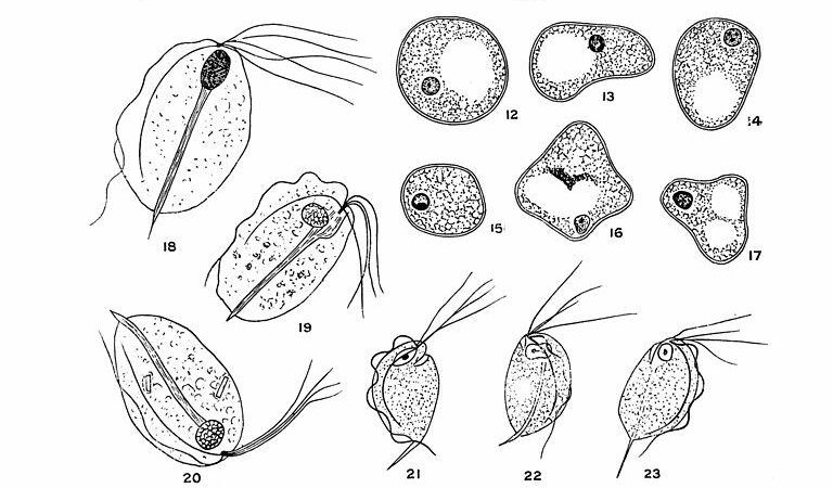
Giardia
Faecal Parasite diagnosis – UKAS accredited test
Faecal microscopy
A concentration technique is routinely performed on all faecal samples for the presence of cysts, ova, and larvae. Direct saline smears are examined on samples that are 24 hours old or less where trophozoite stages of protozoan parasites are suspected.
Sample requirements:
- Minimum of a quarter specimen pot of faecal sample.
- Ova, cysts, and parasites may be passed intermittently so three samples may need to be examined.
- Ova, cysts, and parasites will diminish over time so it is ideal if the sample is less than two days old upon receipt at LSTM. However, older stools will not be rejected.
Key factors affecting test:
Trophozoites may only survive for up to 24 hours in voided faecal samples.
Giardia lamblia (intestinalis) Antigen
If cysts cannot be identified by routine microscopy faecal antigen detection is a sensitive method of diagnosis.
Sample requirements:
- Specimens should be fresh (< 72 hours old).
- OR, sample may be freshly-frozen at -20°C for up to 90 days if the test cannot be performed within 72 hours.
- Minimum quarter pot of faecal specimen.
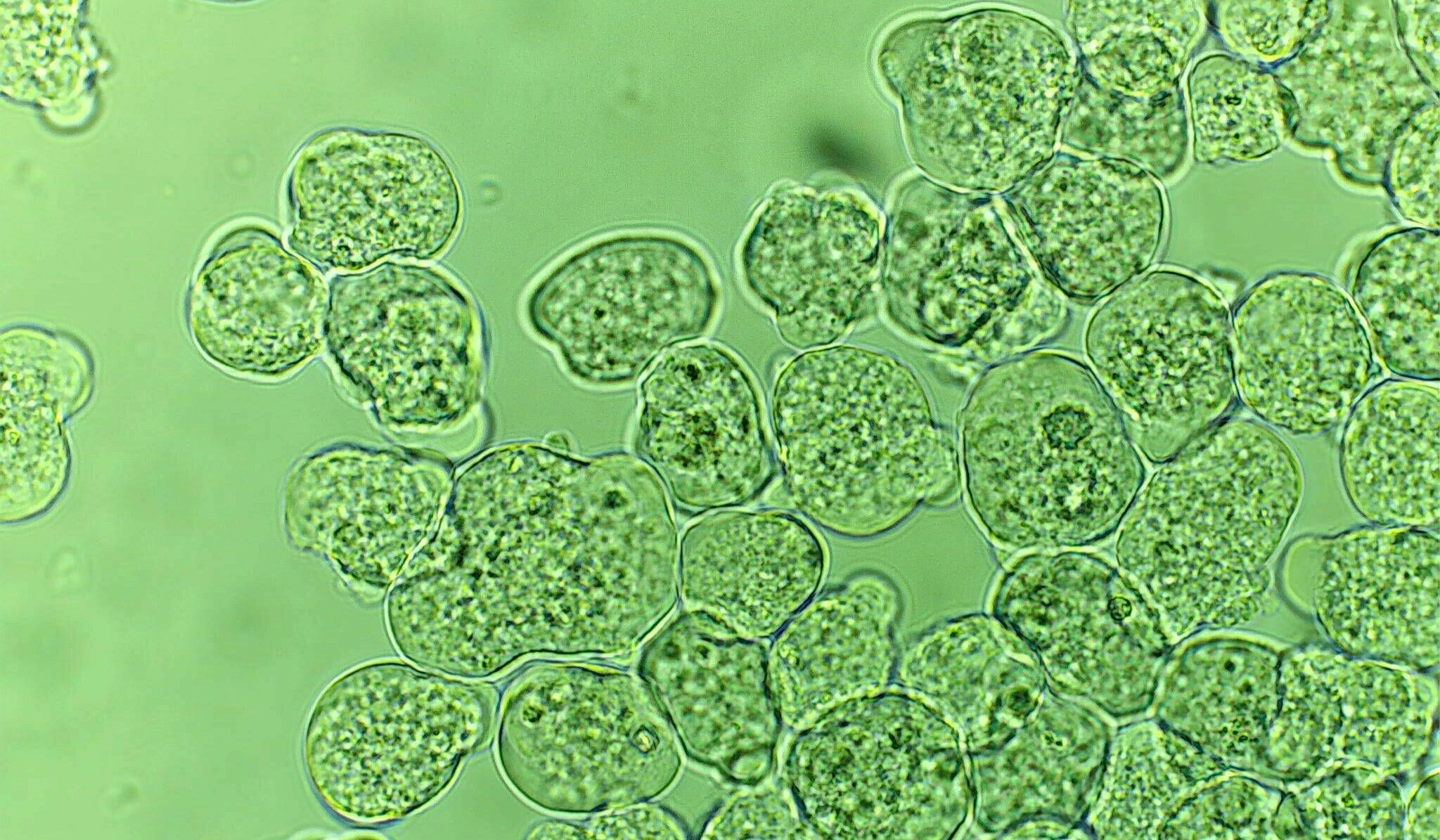
Entamoeba histolytica
Faecal parasite diagnosis – UKAS accredited test
Faecal microscopy (including hot stool)
A concentration technique is routinely performed on all faecal samples for the presence of cysts, ova and larvae. Direct saline smears are examined on samples that are 24 hours old or less where trophozoite stages of protozoan parasites are suspected.
Sample requirements:
- Minimum of a quarter specimen pot of faecal sample.
- Ova, cysts, and parasites may be passed intermittently so three samples may need to be examined.
- Ova, cysts, and parasites will diminish over time so it is ideal if the sample is less than two days old upon receipt at LSTM. However, older stools will not be rejected.
Key factors affecting test:
Trophozoites may only survive for up to 24 hours in voided faecal samples
If Entamoeba histolytica trophozoites are suspected a “hot” stool must be examined.
Hot stool sample requirements:
- Specimen must reach the laboratory within 30 minutes of sample being produced and must be marked as a hot stool, so it can be examined with priority.
- Methanol fixed faecal smears can be made at requesting site if the sample can not arrive at CDPL within 30 minutes. These smears can then be sent to CDPL for staining and examination.
Faecal Antigen detection
Entamoeba histolytica adhesin ELISA
To differentiate between cysts of E.histolytica/dispar as seen by faecal microscopy.
Sample requirements:
- Samples must not be bloody.
- The test must be performed on fresh (< 48 hour old) not fixed specimens
- Unpreserved specimens should be kept at 2-8°C and tested within 48 hours of defaecation.
- If the test cannot be performed within 48hours store sample at -20°C or lower until tested.
- Do NOT send preserved specimens.
- Minimum quarter pot of faecal specimen.
Key factors affecting test:
Test is not validated on samples older than specified.
Amoebiasis – examination of cyst aspirate/pus samples – UKAS accredited test
Specimen requirements:
- Requests for Entamoeba histolytica in samples of pus from liver abscesses should be examined within one hour of collection.
- OR, fresh thin smears of pus should be made (minimum of three), fixed when dry (one minute in methanol) and sent for staining with pus sample.
Key factors affecting test:
About 30 minutes after aspiration of cyst pus/fluid, E.histolytica trophozoites become indistinguishable from macrophages.
Other intestinal protozoa
Faecal Parasite diagnosis – UKAS accredited test
Faecal microscopy (including hot stool)
A concentration technique is routinely performed on all faecal samples for the presence of cysts, ova, and larvae. Direct saline smears are examined on samples that are 24 hours old or less where trophozoite stages of protozoan parasites are suspected.
Sample requirements:
- Minimum of a quarter specimen pot of faecal sample.
- Ova, cysts, and parasites may be passed intermittently so three samples may need to be examined.
- Ova, cysts, and parasites will diminish over time so it is ideal if the sample is less than two days old upon receipt at LSTM. However, older stools will not be rejected.
Key factors affecting test:
Trophozoites may only survive for up to 24 hours in voided faecal samples.
If Entamoeba histolytica trophozoites are suspected a hot stool must be examined.
Hot stool sample requirements:
- Specimen must reach the laboratory within 30 minutes of sample being produced and must be marked as a hot stool, so it can be examined with priority.
- Methanol fixed faecal smears can be made at requesting site if the sample can not arrive at CDPL within 30 minutes. These smears can then be sent to CDPL for staining and examination.
Faecal staining methods
Cryptosporidia
Slides from concentrated faecal deposit are stained using the Z-N technique for identification of oocysts.
Sample requirements:
- Minimum of quarter pot of faecal sample.
- Ova, cysts and parasites may be passed intermittently so three samples may need to be examined.
Intestinal cestodes – tapeworms
Faecal Parasite diagnosis – UKAS accredited test
Faecal microscopy
A concentration technique is routinely performed on all faecal samples for the presence of cysts, ova and larvae.
Sample requirements:
- Minimum of a quarter specimen pot of faecal sample.
- Ova, cysts, and parasites may be passed intermittently so three samples may need to be examined.
- Ova, cysts, and parasites will diminish over time so it is ideal if the sample is less than two days old upon receipt at LSTM. However, older stools will not be rejected.
Whole worms/proglottids
Identification of whole worms/proglottids
Sample requirements:
- Whole worms such as Ascaris may be stored in 10% formol saline. Suspected Taenia species proglottids are best stored in physiological saline and sent to the laboratory as soon as possible.
- The use of 10% formol saline should be avoided unless the sample cannot be sent to the reference laboratory within three days.
Key factors affecting tests:
Identification may be difficult if formalin-fixed proglottids are sent for identification, however proglottids may disintegrate if in saline for three days or more.
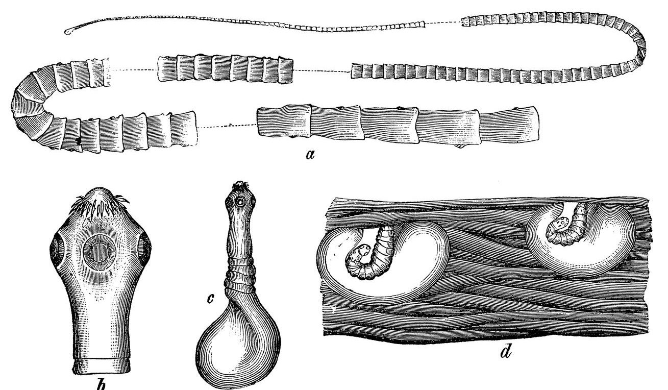
Intestinal trematodes – flukes
Faecal Parasite diagnosis – UKAS accredited test
Faecal microscopy
A concentration technique is routinely performed on all faecal samples for the presence of cysts, ova and larvae.
Sample requirements:
- Minimum of a quarter specimen pot of faecal sample.
- Ova, cysts, and parasites may be passed intermittently so three samples may need to be examined.
- Ova, cysts, and parasites will diminish over time so it is ideal if the sample is less than two days old upon receipt at LSTM. However, older stools will not be rejected.
Sputum
Sputum microscopy is performed for parasitic ova.
Specimen requirements:
- Age and volume of sample is not critical.
- The laboratory does not process samples when a diagnosis of TB cannot be excluded.